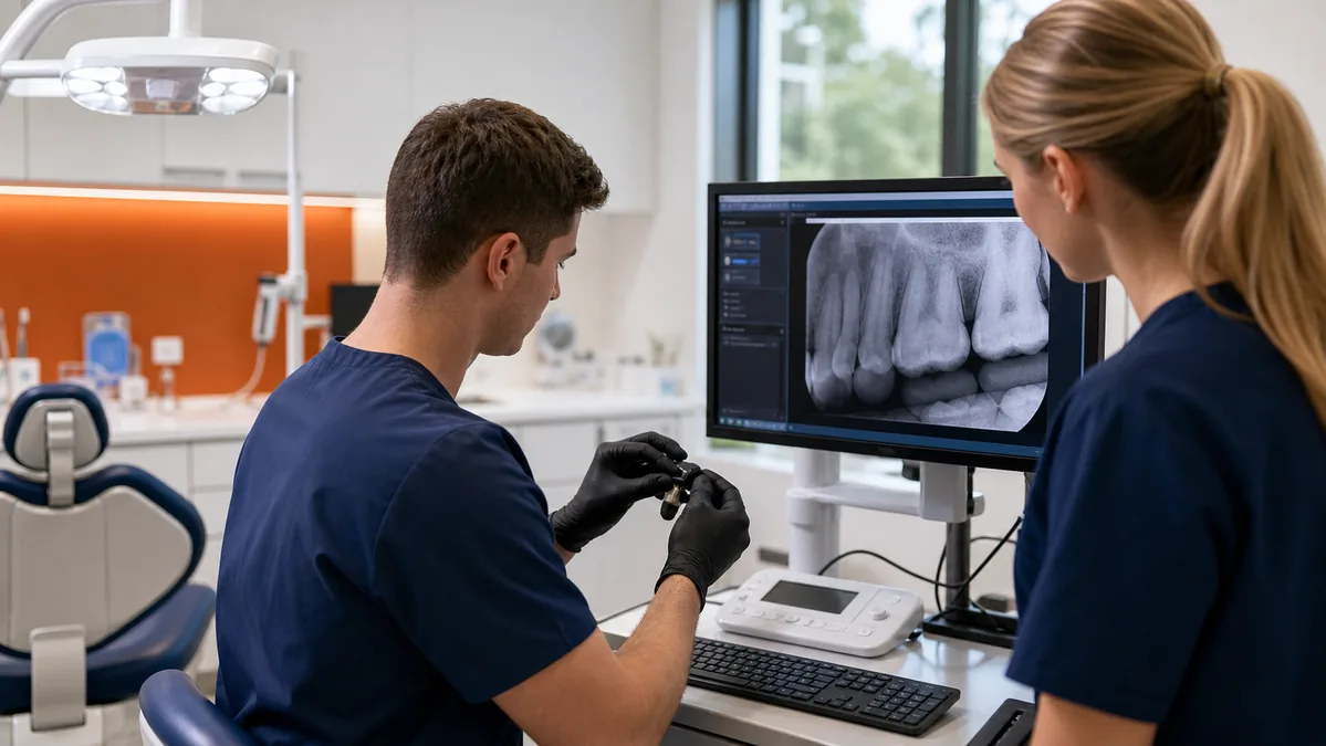
An intraoral sensor does not work in isolation. Image capture depends on the sensor and control unit, approved drivers, acquisition software, workstation, USB or network connection, patient context, storage, practice-management bridge, display, and permissions. A reliable setup validates that whole chain.
Inventory every dependency
Record each sensor and serial number, warranty, operatory assignment, acquisition software and version, driver, workstation, port or network path, imaging database, bridge, practice-management system, and support contact. Include scanners, cameras, panoramic or CBCT equipment, and image-sharing workflows that depend on the same environment.
Confirm compatibility before installation
Verify supported operating systems, hardware, drivers, ports, adapters, and software versions directly with the vendor. Do not assume two products connect because both appear in a marketing list. Confirm whether an official bridge or interface is needed, who supplies it, and who supports each side.
Use a controlled pilot workstation before broad deployment. Keep approved installers and configuration documentation available for recovery, but store software and credentials securely.
Design the workstation and network path
Use stable, business-grade workstations and cabling. Avoid unpowered hubs and improvised extensions where the manufacturer does not support them. For networked imaging, measure throughput, latency, name resolution, storage performance, and segmentation. Wireless can be appropriate for supported workflows, but wired connectivity is often easier to control for fixed clinical stations.
Protect images and patient information
Dental images can contain protected health information. Use unique accounts, role-based access, encrypted transmission where appropriate, secure remote access, device encryption when supported, audit logging, and documented retention and disposal processes. Separate guest traffic and unmanaged devices from clinical systems.
Vendor access should be approved, time-bound where possible, and logged. Remove obsolete remote tools and accounts when a device or support relationship changes.
Back up the complete imaging system
Identify the image database, files, application configuration, license or activation dependencies, and any server components needed for recovery. Protect backup credentials, retain separated copies, monitor failures, and test restoration to a nonproduction environment. Confirm that restored images reconnect to the correct patient context.
Run a clinical workflow test
- Open the correct patient and operatory workflow.
- Acquire a test image using an approved process.
- Confirm orientation, display, storage, and retrieval.
- Open the image through the practice-management bridge.
- Validate access from all intended clinical stations.
- Test export, referral, or sharing workflows without exposing real patient data.
- Document results, versions, and the support path.
Troubleshoot from device to database
When capture fails, record the exact error, affected devices and operatories, recent changes, whether another sensor works on the same station, and whether the sensor works on a known-good station if the vendor permits that test. This helps separate device, driver, workstation, network, application, storage, and bridge causes.
Security references
Always follow the current manufacturer and software-vendor instructions for the exact device and version in use. This guide is not clinical, legal, or manufacturer service advice.
Related resources